W. Scott Young, M.D., Ph.D.
Brief Research Summary Below and Selected Bibliography Here
Scientist Emeritus, NIMH, 2022- | |
Principal Investigator, retired 2022 |
 |
|
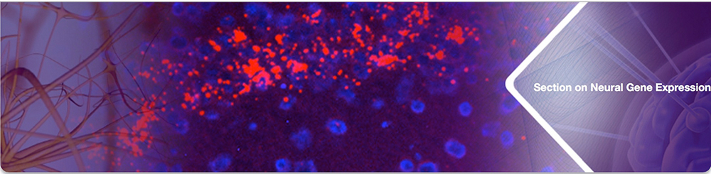
The Section on Neural Gene Expression at the National Institute of Health in Bethesda, Maryland, USA, investigated the roles and regulation of expression of vasopressin (Avp) and oxytocin (Oxt) in the central nervous system. They are peptide hormones composed of 9 amino acids and that participate in the regulation of fluid balance, parturition and lactation. In addition, they have important roles in various behaviors, including social and maternal ones, through their actions mediated by at least 3 receptors in the brain. Our group used a variety of techniques, including anatomical (hybridization histochemistry and receptor autoradiography), molecular biological, transgenic animals, optogenetics, and electrophysiology to explore behavior in the mouse. For example, we generated mice lacking functional Oxt, as well as mice that express green fluorescent protein in Oxt neurons, in our attempts to determine the essential and non-essential roles of this hormone. We also created the first conditional knockout of the oxytocin receptor (Oxtr) and used them to further our knowledge of Oxt's role in behavior. For example, we showed that the Oxtr is necessary for intra-strain, but not inter-strain, social recognition. Inactivation of the Oxtr in forebrain excitatory neurons decreases fear conditioning. Inactivation of the Oxtr in serotonin neurons in the brainstem reduces agression in males, but not females, without affecting anxiety-like behaviors. Our last work examined the role of the vasopressin 1b receptor (Avpr1b) in the brain. It is found predominantly in the CA2 region of the hippocampus, an area that receives innervation from the AVP-producing paraventricular nucleus of the hypothalamus. We made Avpr1b knockout mice to inactivate the receptor and observed that these mice have a marked reduction in social, but not predatory or defensive, aggression in males and females. They also have modest declines in social recognition in males and females. We thus proposed in 2006 that the CA2 region is necessary for proper social memory and aggression. We subsequently confirmed this in further studies. We showed that viral replacement of Avpr1b in the CA2 area of the mouse hippocampus in these knockout mice restores aggression. Furthermore, the Avpr1b (and Oxtr) enables significant potentiation of excitatory synaptic responses in CA2, but not in CA1 or in slices from Avpr1b (and Oxtr, respectively) knockout mice. Next, we demonstrated that optogenetic stimulation of vasopressin fibers within the mouse CA2 strongly enhances social, but not object memory. This vasopressin release from fibers arising in the paraventricular nucleus lengthens the social memory by at least 80 fold and is inhibited by a locally infused Avpr1b antagonist. In addition, the enhancement is produced by stimulation during the acquisition phase of the memory and not during the recall phase. We also demonstrated that encoding of social recognition memory in the CA2 region entails suppression of pyramidal neurons’ activity leading to a sparse representation of the familiar conspecific. Interestingly, our research shows that facilitation of social memory and aggression by the CA2 regions does not require the NMDA NR1 receptor subunit. My last research talk at NIMH is available. I retired in 2022 to become Scientist Emeritus. Here are some further insights. While a Neurology Resident at the University of Virginia, I also performed some basic research (1981-1982) in the laboratory of Prof. Lennart Heimer on the ventral pallidal projection to the mediodorsal thalamus. | |
Department of Pharmacology and Experimental Therapeutics, | |
Ph.D. Graduate StudentI received my B.A., M.D. and Ph.D from The Johns Hopkins University. The latter degree, obtained under the guidance of Prof. Michael Kuhar, described the development of in vitro receptor autoradiography and the first applications of the technique to the anatomical localization of neurotranmsitter receptors in human and other animal brains. We developed the first autoradiogram showing the anatomical locations of the receptor for a diffusable ligand on September 26, 1978.
As an undergraduate, I assisted Dr. Milton Saier in Prof. Saul Roseman's lab and then examined the development of chloramphenicol glucuronyl transferase in Prof. Paul Lietman's lab as a medical student.
| |